
How Fitzpatrick Phototype Changes Treatment Decisions in Aesthetic Practice
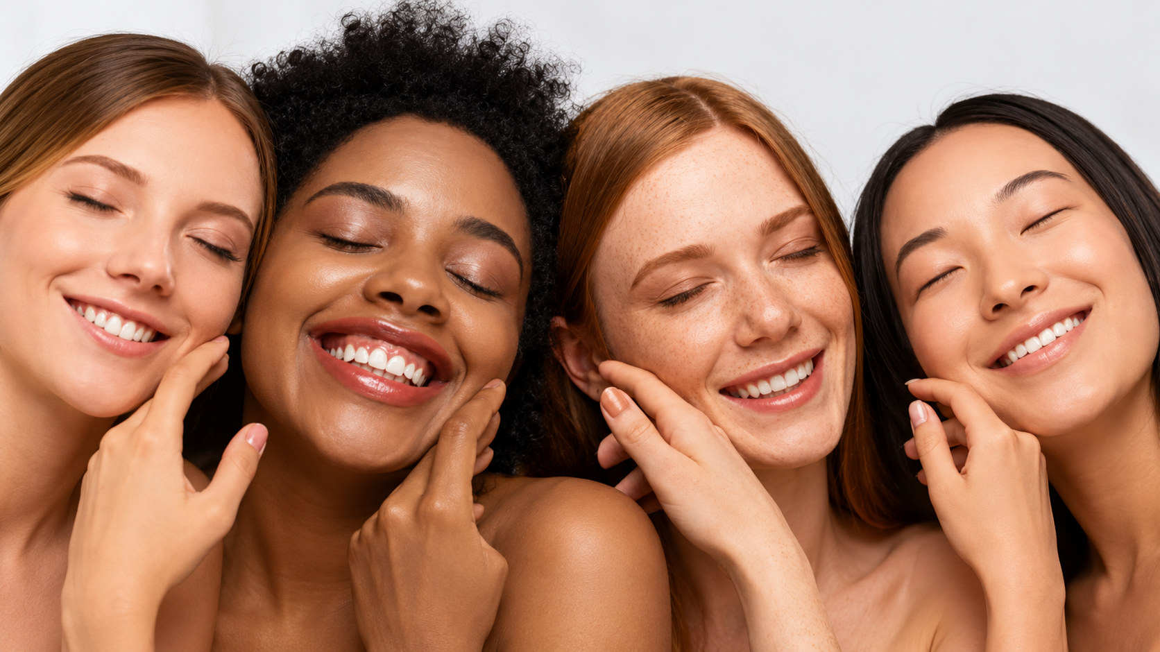
For a long time, aesthetic protocols were written around one kind of skin and applied to everyone. Settings, peel strengths, and treatment intervals were built from clinical experience with lighter phototypes, and patients who responded differently were treated as complications rather than as predictable variation.
That approach produces a specific and avoidable outcome, which is post-inflammatory hyperpigmentation in patients who were never assessed for their risk of it. For a licensed esthetician or an Aesthetic Medical Assistant (AMA), understanding how phototype changes the calculation is one of the more directly useful pieces of clinical knowledge available.
What Fitzpatrick actually measures
The Fitzpatrick scale classifies skin by how it responds to ultraviolet exposure, from Type I, which burns and does not tan, through Type VI, which rarely burns and tans deeply. It was developed in the 1970s to help dose ultraviolet phototherapy, and that origin explains both its usefulness and its limits.
It is a clinical classification, not a description of ethnicity. Patients from any background can fall anywhere on the scale, and treating phototype as a proxy for heritage produces both clinical errors and uncomfortable conversations. The correct question at intake is how the skin behaves in the sun, not where the patient’s family is from.
The scale also has real weaknesses. It relies on self-report, and patients are frequently inaccurate about their own burning and tanning history. It correlates only loosely with pigmentation risk at the higher end, where Types V and VI cover a very wide range of actual melanin content and reactivity. Used alone it is a starting point, not an assessment.
Why pigmentation risk changes with phototype
Melanocyte density is broadly similar across phototypes. What differs is melanin output, the size and distribution of melanosomes, and how readily melanocytes respond to a stimulus. In more pigmented skin, melanosomes are larger, more heavily melanized, and distributed individually through the epidermis rather than clustered, and the melanocytes themselves are more reactive to inflammatory signaling.
The practical consequence is that any inflammatory event, including one that was clinically appropriate and well executed, can trigger pigment production that outlasts the original insult by months. This is why post-inflammatory hyperpigmentation is the dominant complication in higher phototypes while erythema and vascular reactivity dominate in lower ones.
There are structural differences beyond pigment as well. Stratum corneum cohesion tends to be greater in more pigmented skin, dermal fibroblasts are often larger and more numerous, and the tendency toward hypertrophic scarring and keloid formation is higher. Each of these affects how a treatment behaves.

Where phototype changes the protocol most
Light-based treatments carry the largest differential. Intense pulsed light targets melanin as a chromophore, which means that in higher phototypes the epidermal pigment competes with the intended target and absorbs energy meant for the vessel or hair follicle. The result can be burning, blistering, and lasting pigment change. Longer wavelength devices with longer pulse durations and aggressive epidermal cooling are the safer approach, and device selection here is a physician decision with real consequences.
Chemical peels are workable across phototypes but the depth calculation changes. Superficial peels are generally well tolerated. Medium and deep peels carry substantially higher pigmentation risk in Types IV through VI, and pre-treatment with a pigment-suppressing agent, along with strict photoprotection before and after, becomes part of the protocol rather than an optional extra.
Microneedling is comparatively forgiving, because it works mechanically rather than through selective absorption of light by pigment. It has become a preferred modality in higher phototypes for exactly that reason, though depth and pass count still need restraint, since the risk driver is inflammation rather than the mechanism itself.
Laser hair removal follows the same logic as other light-based treatments. Wavelength selection and cooling do most of the safety work, and settings appropriate for one phototype can cause injury in another.
What to establish at intake
A short set of questions surfaces most of the risk. Ask whether the patient has ever developed dark marks after a cut, a burn, acne, or a previous treatment, because a personal history of post-inflammatory hyperpigmentation is a stronger predictor than phototype alone. Ask about melasma, which behaves differently from ordinary PIH and can be aggravated by heat as well as by light. Ask about keloids or raised scars.
Then establish the current situation. Recent sun exposure or a tan changes how the skin will absorb energy on the day. Ongoing use of hydroquinone or other pigment-suppressing agents matters. Photoprotection habits predict whether any result will hold. Recent isotretinoin and current pregnancy both change what is appropriate.
Document the answers rather than remembering them. A photograph in consistent lighting at baseline is worth more than any note when a question arises three weeks later.
What goes wrong in practice
Most pigmentation complications trace back to one of four things. Settings chosen from a default protocol rather than from an assessment. No test patch in a patient whose response was uncertain. No pre-treatment or photoprotection plan in a patient who needed one. Or a treatment interval too short for inflammation from the previous session to have resolved.
None of these are exotic failures. They are ordinary shortcuts, and they are the reason patients with higher phototypes have historically been undertreated by cautious clinics and injured by careless ones. Neither outcome is acceptable, and the middle path is assessment rather than avoidance.
Scope and the assistant’s role
Determining phototype, selecting a device, choosing settings, and deciding whether to treat are all clinical decisions that belong to the supervising physician or the appropriately licensed provider.
What clinical support staff contribute is substantial regardless. Taking a complete pigmentation and scarring history rather than a summary. Capturing consistent baseline photography. Noticing that a patient has a fresh tan before the room is set up rather than after. Reinforcing photoprotection instructions in language the patient will actually follow. Flagging a response at follow-up that does not match what was expected.
If there are topics you would like to see covered next, write to us at editorial@upsthetics.com.
Disclaimer
This content is educational only. It does not constitute medical advice and does not qualify anyone to perform aesthetic or medical procedures. Scope of practice and licensing requirements vary by state, and every professional is responsible for working within their own license.
FAQS
Is Fitzpatrick skin type the same as ethnicity?
No. The Fitzpatrick Skin Phototype Classification measures how an individual’s skin responds to ultraviolet (UV) radiation, particularly its tendency to burn or tan after sun exposure. It is not a measure of ethnicity, race, or skin color alone. People from the same ethnic background may have different Fitzpatrick skin types, so phototype should always be determined through clinical history rather than appearance.
Why is post-inflammatory hyperpigmentation (PIH) more common in darker skin?
Individuals with higher Fitzpatrick phototypes have more active melanocytes and produce larger, more heavily pigmented melanosomes. Following inflammation or skin injury, these cells may generate excess melanin, increasing the likelihood of post-inflammatory hyperpigmentation (PIH). Because PIH can persist for months, minimizing unnecessary inflammation and selecting appropriate treatment parameters are important clinical considerations.
Can patients with darker skin safely undergo laser treatments?
Yes. Many laser procedures can be performed safely in patients with darker skin when the appropriate technology, wavelengths, pulse durations, cooling methods, and treatment settings are selected. Careful patient assessment and device selection help reduce the risk of burns, dyspigmentation, and other complications. Treatment planning should always be performed by appropriately qualified healthcare professionals.
Is a test patch always recommended before laser treatment?
A test patch is not required for every patient, but it is commonly recommended whenever the clinical response is uncertain. It is particularly valuable when treating higher Fitzpatrick phototypes, using new devices, treating previously untested areas, or managing patients with a history of post-inflammatory hyperpigmentation or pigmentary complications. Test patches can help evaluate tissue response before proceeding with full treatment.
Why is Fitzpatrick skin typing important in aesthetic medicine?
The Fitzpatrick classification helps practitioners evaluate how the skin may respond to UV exposure and certain aesthetic procedures. It is widely used during treatment planning to guide clinical decision-making, support patient safety, and reduce the risk of complications such as burns, post-inflammatory hyperpigmentation, or hypopigmentation, particularly when performing laser and light-based treatments.
Stay Updated
Related posts

The Skin Microbiome and Why Some Patients Tolerate Treatments Poorly
Smoking and skin aging: what aesthetic professionals and patients should know
Skin aging by decade: what changes and what is commonly discussed in aesthetic practice
About the author
